
- Home
- Procedures
- Resection of benign and malignant tumours in Tijuana
Resection of benign and malignant tumours in Tijuana
Removal of soft-tissue, abdominal wall and digestive organ tumours, with adequate margins and histopathological study. Diagnosis first, surgery afterwards.
The correct sequence: diagnosis, staging, surgery
The costliest error in cancer surgery is operating before knowing. A suspicious lesion excised without planning can be left with insufficient margins, seed the track and force a second, wider operation on an already scarred field. That complicates everything that follows.
The correct sequence is the opposite: first the lesion is characterised with imaging — ultrasound, CT or MRI according to its location — then a histological diagnosis is obtained by biopsy where indicated, and only then is the resection planned with the margin that diagnosis demands. In small, clearly benign lesions, excisional biopsy resolves both stages in a single act.
Which tumours are resected here
Within general surgery, benign and malignant tumours of soft tissue, of the abdominal wall, of the thyroid gland and of the digestive organs are resected: stomach, small bowel, colon, rectum, liver and gallbladder.
Each location has its own logic of margin and staging. In a lipoma it is enough to excise it intact. In a colonic tumour the segment must be resected with its vascular and lymphatic territory. In a thyroid tumour, the extent of thyroidectomy is defined by the prior cytology. There is no single technique: there is a single principle, which is to remove all the tumour with the margin its biology demands.
The work does not end in theatre
The resected specimen is oriented and sent to the histopathology laboratory, which reports the tumour type, the grade, the status of the margins and, where applicable, the number of nodes examined and involved. That report is what defines whether the operation was sufficient or whether more treatment is needed.
When the result is malignant, the next step is coordination with medical oncology and radiotherapy. Surgery is one part of the treatment, not always the only one, and the patient should leave the consultation knowing the complete plan, not only what happened in theatre.
What the procedure involves
When it is indicated
- Soft-tissue tumour larger than five centimetres or growing rapidly
- Abdominal wall tumour, benign or malignant
- Thyroid cancer confirmed by cytology
- Digestive tract tumours with a surgical indication
- A lesion with a malignant histological diagnosis requiring wider margins
- Symptomatic benign tumour, or one with functional or cosmetic impact
Techniques and resources
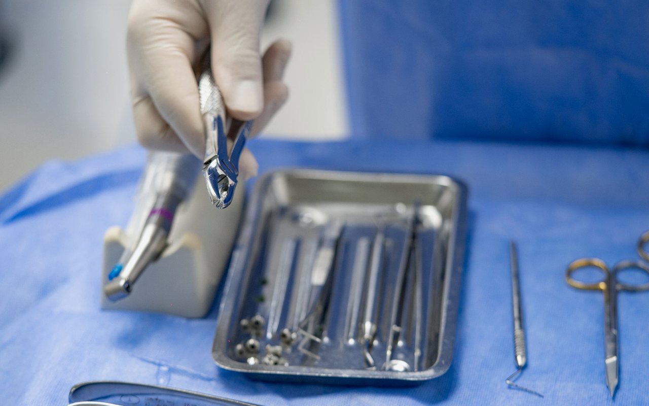
- Cancer surgery instrument set
- Laparoscopic tower
- Advanced energy sealing devices
- Marking and orientation of the surgical specimen
- Intraoperative frozen section where the case requires
Frequently asked questions about tumor resection
Because in suspicious lesions that sequence can prove expensive. If it turns out malignant and was excised without a margin, reoperation on already scarred tissue is needed, with more surgery and a worse result. In small, clearly benign lesions it is done in one act, because complete excision is at the same time the biopsy.
No. The word tumour means a mass or growth, and the great majority seen in clinic are benign: lipomas, cysts, fibromas, thyroid nodules. Histopathological study is what separates one from the other, which is why every specimen goes to pathology.
It depends on the type, grade and stage. In some tumours complete resection is the definitive treatment; in others chemotherapy, radiotherapy or both are needed, before or after surgery. That plan is defined with oncology and explained to you in full, not in instalments.
Bring your studies and settle the question
One consultation with your ultrasound or CT in hand is worth more than ten internet searches. Book by phone or through the form and you will be told what is there, what it means and what your options are.